animal cell electron microscope
An animal cell also contains a cell membrane to keep all the organelles. Animal cell under electron.

Microscopic Animal Cells Images Kuhn Photo
Animal cell under electron microscope.

. An animal cell also contains a cell membrane to keep all the organelles and cytoplasm contained but it lacks a cell wall. Two types of electron microscope have been. Two types of electron microscopytransmission and scanningare widely used to study cells.
A typical animal cell as seen in an electron microscope Medical Images For PowerPoint 1. Animal cells have a basic structure. A typical animal cell is 1020 μm in diameter which is about one-fifth the size of the smallest particle visible to the naked eye.
Plant animal and bacterial cells have smaller components each with a specific function. An animal cell also contains a cell membrane to keep all the organelles and cytoplasm contained but it lacks a cell wall. Electron Microscopic Study Of Cell And Organelles Important The largest known animal cell is.
Observing a wide range of biological processes and animal cell under light microscope is easier due to advances in microscopic techniques. The animal cell is more fluid or elastic or malleable in structure. Select from premium Animal Cell Microscope of the highest quality.
Below the basic structure is shown in the same animal cell on the left viewed with the light microscope and on the right with the transmission electron. In principle transmission electron microscopy is similar to the observation of stained cells with. What microscope is used to view animal and plant.
A membrane that is transparent to electrons protects the fully hydrated sample. Animal cells do not have a cell wall. As a result most animal cells are round and flexible whereas most plant cells are rectangular and rigid.
Plant cells are rectangular in shape and larger than animal cells. Animal Cell In Electron Microscope. Electron Microscopic Study Of Cell And Organelles Important The largest known animal cell is the ostrich egg which can stretch.
What can be seen with an electron. How does electron microscope affect cells. Animal cell under electron microscope.
Animal Cell Under An Electron Microscope. What microscope is used to view animal and plant. Beneath a plant cells cell wall is a cell membrane.
Below the basic structure is shown in the same animal cell on the left viewed with the light microscope and on the right with the transmission electron. Light and electron microscopes allow us to see inside cells. Electron microscopes are the most powerful type of microscope capable of distinguishing even individual atoms.
Structure Of Plant And Animal Cells Under An Electron Microscope Ppt Video Online Download

File Anatomy And Physiology Of Animals Animal Cell Electron Microscope Jpg Wikimedia Commons
Chapter 17 Opercularia 01 Ope3847 Fig 1 Opercularia Coarctata Cross Section Of Cell

Gulf War Illness Not In Veterans Heads But In Their Mitochondria Scienceblog Com Animal Cell Organelles Mitochondria Biotechnology Art

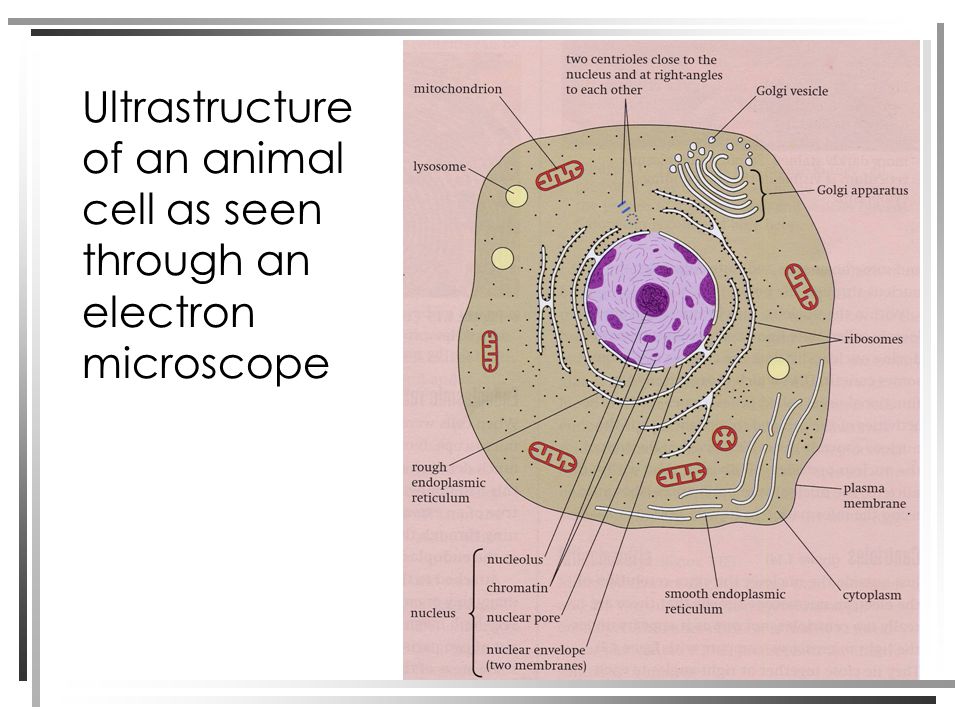
Ultrastructure

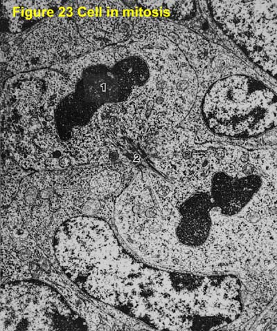
1 2 Skill Interpretation Of Electron Micrographs Youtube
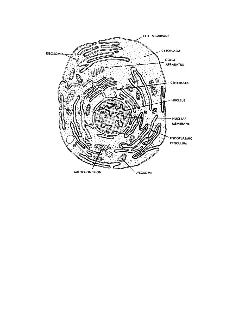
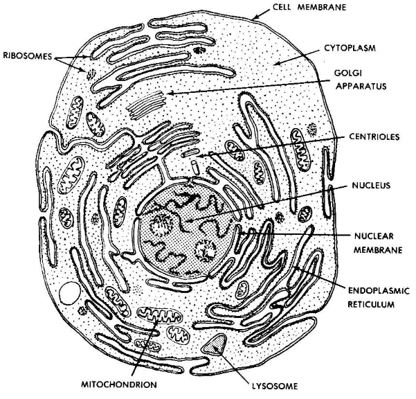
Major Components Of A Typical Animal Cell Basic Human Anatomy

Illustrate A Plant Cell As Seen Under Electron Microscope How Is It Different From Animal Cell Science The Fundamental Unit Of Life 16643377 Meritnation Com

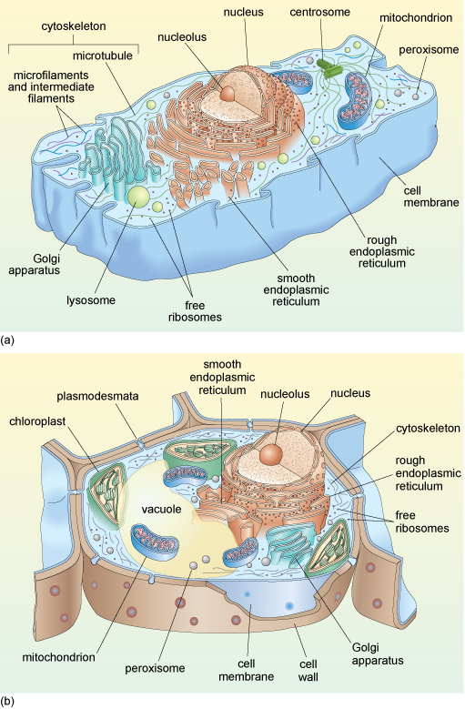
Animal Plant Cells 1 2 2 Cie A Level Biology Revision Notes 2022 Save My Exams
Images 01 Introduction And Terminology Basic Human Anatomy

Cells Under Electron Microscope Google Search Animal Cell Structure Cell Diagram Animal Cell
![]()
Electron Micrograph Animal Cell Hi Res Stock Photography And Images Alamy

Draw A Large Diagram Of An Animal Cell As Seen Through An Electron Microscope Label The Parts That Ca Brainly In

Aice Biology Chapter 1 Animal Cell Electron Micrograph Labeling Diagram Quizlet
![]()
Transmission Electron Micrograph Tem Of Lysosomes A Lysosome Is A Membrane Bound Organelle Found In Nearly All Animal Cells They Are Spherical Vesicles Which Contain Hydrolytic Enzymes That Can Break Down Virtually All
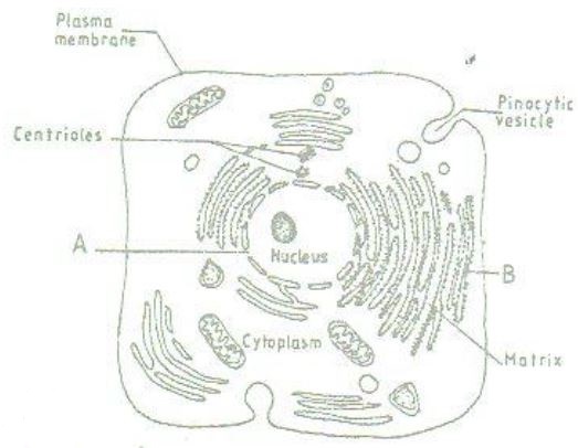
The Figure Below Is A Fine Structure Of A Generalized Animal Cell As Seen Under An Electron Tutorke

The Animal Cell Sahani Perera Bulb
A Tour Of The Cell View As Single Page
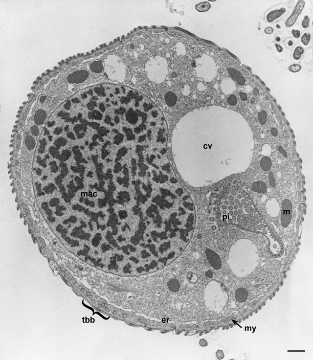
Electron Micrographs